Ultrasound of fetal central nervous system
Fetal neurosonography – detailed ultrasound examination of the fetal central nervous system.
Fetal neurosonography is a detailed ultrasound examination of the fetal brain, spine, and spinal cord. It is a comprehensive diagnostic examination used when it is necessary to assess fetal central nervous system development in greater detail or to evaluate potential signs of brain injury.
The examination is not limited to measuring the fetal brain. During neurosonography, the structure of the fetal brain, the brain midline, lateral ventricles, cerebellum, brainstem, cortical development, and, when needed, cerebral blood flow are assessed. The examination also includes assessment of the fetal spine and spinal cord. If spina bifida is suspected, both the spinal finding and the associated changes in the brain are evaluated.
The aim of fetal neurosonography is to help determine whether the visible development of the fetal brain, spine, and spinal cord corresponds to the gestational age, whether a prior suspicion is confirmed, and whether further investigations or involvement of other specialists is needed.
When is fetal neurosonography recommended?
Fetal neurosonography may be recommended when a previous ultrasound examination has shown an unclear or suspicious finding in the area of the fetal brain, spine, or spinal cord, or when the pregnancy is associated with an increased risk of fetal central nervous system injury.
The examination may be needed, for example, if the fetal brain ventricles are enlarged, if a cyst or a suspected cyst is seen in the fetal brain, if the fetal head measurements do not correspond to the gestational age, or if a previous examination has raised suspicion of a developmental abnormality of the brain, spine, or spinal cord.
Neurosonography may also be recommended if there is a history of central nervous system malformations, psychomotor developmental delay, or intellectual disability in the family or in a previous pregnancy.
The examination may be useful in higher-risk pregnancies, for example, in monochorionic twin pregnancy, severe fetal growth restriction, fetal heart defects, or other congenital malformations. In cases of fetal heart defects or severe growth restriction, a detailed assessment of the brain may be important, as fetal brain development and cerebral blood flow may require special attention.
Neurosonography is also used when intrauterine infection is suspected, for example, cytomegalovirus (CMV) or toxoplasmosis, to assess whether the infection has affected the fetal brain or other organs.
The examination may also be needed if the fetus has abnormalities in other organ systems, such as kidney malformations, limb deformities, clubfoot, cleft lip, or an abdominal wall defect.
None of these findings automatically means that the fetus has a central nervous system injury. The purpose of neurosonography is to determine whether the visible development of the brain, spine, and spinal cord is normal or requires further follow-up, amniocentesis, genetic testing, fetal brain MRI, or involvement of other specialists.
When is the examination performed?
At the Fetal Ultrasound Center, neurosonography is performed primarily during two key time periods.
Early neurosonography at 16–17 weeks of pregnancy helps to clarify unclear findings detected during the 12–13-week scan. The examination may also be important when an intrauterine infection, such as cytomegalovirus (CMV), is suspected during pregnancy, because some infection-related changes may be visible in the fetal brain or other organs. At this gestational age, it is possible to assess the fetal brain midline, lateral ventricles, developing midline structures, posterior fossa, cerebellar region, brainstem, and spine.
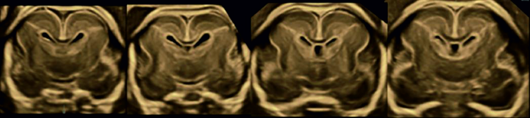
Detailed neurosonography at 20–21 weeks of pregnancy is the most comprehensive ultrasound-based assessment of the fetal central nervous system in mid-pregnancy. The examination assesses the fetal brain, spine, and spinal cord, including the brain midline, the width and internal structure of the lateral ventricles, development of the deep brain nuclei, white matter texture, cerebellum, brainstem, cortical development, and formation of the brain gyri according to gestational age.
Neurosonography is not based only on a subjective impression. During the examination, different brain structures are measured and assessed, their development is compared with gestational-age-specific reference values, and, when needed, fetal cerebral blood flow is also evaluated.
In addition, fetal organ structures are assessed as a whole because the significance of a central nervous system finding often depends on whether it is isolated or associated with developmental abnormalities in other organs.
How is the examination performed?
Fetal neurosonography may require both transabdominal and transvaginal ultrasound.
Transabdominal ultrasound provides an overview of the fetal position, the main brain structures, the spine, and the overall fetal anatomy. High-resolution transvaginal ultrasound may provide a more detailed view of the fetal brain midline, corpus callosum, posterior fossa, cerebellum, and brainstem.
If fetal neurosonography is performed because of a suspected developmental abnormality, the use of high-resolution transvaginal ultrasound is always considered. It may be necessary in order to assess the fetal brain and central nervous system structures as accurately as possible, especially the brain midline, corpus callosum, posterior fossa, cerebellum, and brainstem.
Transvaginal ultrasound is used only when medically necessary and with the patient’s consent. Its purpose is to obtain the most accurate possible diagnostic information about the fetal brain, spine, and spinal cord.
What can the examination help decide?
Neurosonography helps clarify whether a prior suspicion of a developmental abnormality of the fetal brain, spine, or spinal cord is confirmed. It also helps to assess whether the finding requires further follow-up, amniocentesis, genetic testing, fetal brain MRI, or involvement of other specialists.
Fetal neurosonography is the basic examination for assessing the fetal central nervous system in prenatal diagnostics. In some cases, well-performed neurosonography may provide as much, or even more, practical diagnostic information than fetal brain MRI. Therefore, not every neurosonographic finding automatically requires confirmation by MRI.
Fetal brain MRI does not replace neurosonography; rather, it complements it in selected cases. MRI may be needed when, after a detailed neurosonographic examination, a specific question remains that ultrasound cannot sufficiently answer. In such cases, neurosonography helps to formulate the precise question that MRI should answer.
The aim of neurosonography is not only to confirm a diagnosis. Equally important is counseling the family about what the detected developmental abnormality may mean for the child’s life, health, and development, as well as possible treatment or follow-up after birth.
Neurosonography is a very detailed structural examination, but it cannot fully predict a child’s future learning ability, speech development, behavior or all neurological functions. The value of the examination lies in assessing, as accurately as possible, the visible development of the fetal brain, spine, and spinal cord, counseling the family about the possible significance of the finding, and planning further investigations, pregnancy follow-up, and postnatal care when needed.
When necessary, neurosonography can also help plan consultation with a clinical geneticist, pediatric neurologist, neonatologist, neurosurgeon, or another specialist.