Fetal anatomy screening at 20-22 weeks of gestation
Fetal anomaly scan is one of the most essential ultrasound examinations during pregnancy, performed using an abdominal probe during the 20th-21st week of pregnancy. During the ultrasound examination within 60 minutes, the organ structures of the fetus are checked from the “top-to-toe” to discover possible visible deviations from its normal development. Special attention is paid to the assessment of fetal brain and heart structures.
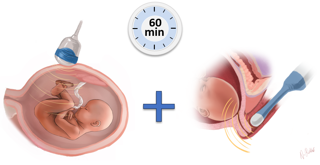
As part of the ultrasound examination of fetal developmental defects, it is possible an assessment of the risk of premature birth and/or to carry out preeclampsia screening as an additional service if desired.
Single pregnancy:
225.00 €
Multiple pregnancy:
275.00 €