3D/4D ultrasound scan
3D scan shows a fetus on the screen as three-dimensional in real life. 4D test enables to see the child’s movements, yarning, smile and sucking of a finger as three-dimensional in real time. The given ultrasound test also applies colour doppler trial which enables to see the blood circulation of the working heart, fetal breathing movements and assess the welfare condition of the fetus.
The fetal 3D/4D ultrasound scan includes:
- Creating of the three-dimensional image of the face and limbs of the fetus;
- Assessment of the position and structure of the placenta;
- Assessment of the quantity of amniotic fluid;
- Assessment of the fetal structures;
- Assessment of the fetal functions (movements, breathing, heartbeat, swallowing);
- If required, assessment of the fetal sex.
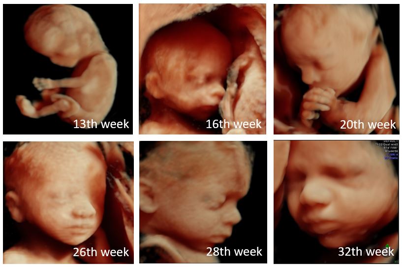
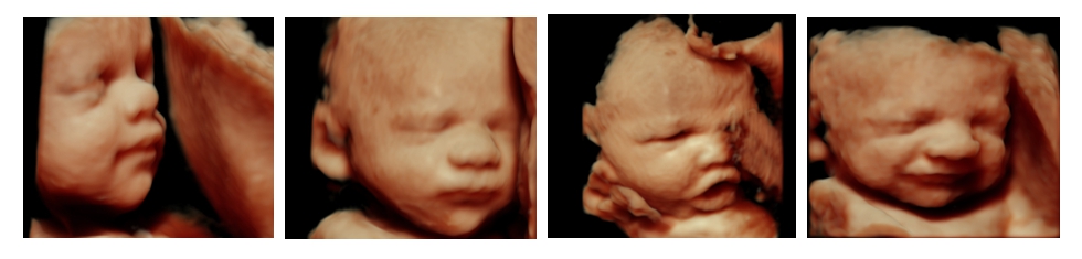
3D/4D ultrasound scan can be performed in any stage of pregnancy. It should be considered that the smaller the pregnancy, the bigger the possibility to see the fetus as a whole. The bigger the pregnancy, the more developed are the mimic muscles of the fetal face, i.e. the more “cute“ and natural is the child’s face. Unfortunately we are not able to see the face of the fetus always. Usually we recommend to come to the ultrasound scan during the pregnancy week 26-32. By that time the fetus has all vital organs developed and the period of rapid growth and maturing continues. The fetus is similar to already newborn infant as to the proportions of its body. It has bigger head compared to its body and thus most of them have already turned themselves upside down. It has developed the mimic muscles of the face and can smile. It opens its eyes. The connection has been created between the eye retina and brain and the child has a characteristic glance in its eyes. The pineal gland of the brain has started to produce the hormone called melatonin. Upon the impact of the given hormone the fetus will create its daily rhythm. Some fetuses are more active in the mornings, others in the evenings, but regardless of the latter the fetuses still sleep most of the time, i.e. 90-95% of the day.
In the given stage of pregnancy the fetus has already all 5 senses developed
- It sees the light gleaming through the mother’s stomach covers
- It is able to hear the sounds coming from outside the womb, e.g. father’s voice
- It breathes and swallows amniotic fluid by feeling its smell and taste
- It has developed skin sensibility to touch and pain
The fetus has, just as the newborn infant, in the given stage of pregnancy already eyebrows and eyelashes. The nails are covering the toe and finger tips. Some fetuses have already hair. Its heart beats 120-160 times a minute. This is twice as fast as its mother’s, i.e. the heartbeat of the mother and father in total. In the course of fetal ultrasound the fetal position is assessed, whether the fetus is upside down or in the breech state. The size, maturity and location of the placenta as to the cervix are examined. It is important to specify that the placenta or its blood vessels would not cover the internal estuary of the cervix. The latter for the purpose that the child could be born.
In the course of ultrasound test the amount of amniotic fluid is assessed, so that it would not be too much or too little. The amniotic fluid enables the fetus to move itself and as the fetus also breaths and swallows amniotic fluid, the fetal lungs and intestines are also developing.
By measuring the size of the skull, the perimeter of the stomach and length of the thighbone it is possible to assess the fetal growth and estimate the approximate birth weight of the child, by knowing that the fetus gains 250-500 g with 2 weeks in this stage of pregnancy, i.e. maximum 1 kilogram with a month. These future mothers who have not gained enough weight by this stage of pregnancy and whose fat of the stomach is not compliant with the size of pregnancy gain most from the assessment of fetal growth.
The welfare condition of the fetus is assessed by using the ultrasound doppler trial in the course of which the blood flow indices of umbilical cord artery of the fetus is assessed.
If required, and depending on the position of the fetus, the following can be additionally required:
- Ultrasound scan recording on a memory stick including comments;
- Coloured photo-quality picture of the fetus 10x15 cm.
Some tips before the ultrasonography
It would be good if the expecting mother will drink plenty of water - it is recommended to drink 1.5-2.0 l of water a day two days beforehand. The urinary bladder should be emptied and the navel piercing should be removed before the ultrasonography. It is also useful that the gels that contain magnesium are used on belly – the consistency of the ultrasonic gel changes very liquid when exposed to magnesium and shall not stay on the woman’s belly.