Successful 3D/4D ultrasound picture
Most of the patients having registered to 3D/4D test presume that they will leave with a beautiful picture of the child’s face after ultrasound test. Unfortunately, it is not always possible to get a nice 3D picture of the fetal face. The smaller the fetus, the greater the possibility to get a picture of the fetus as a whole.
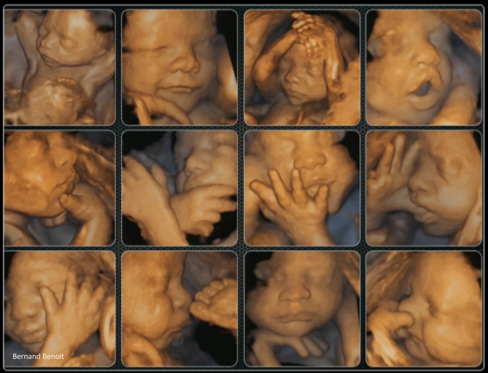
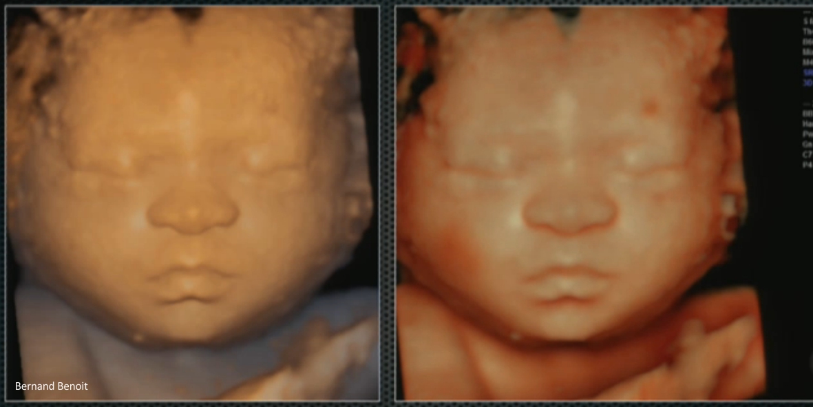
Starting from the 20th pregnancy week the fetus cannot be any more visualized as a whole, but the possibility to visualize the fetal face is greater. The bigger the pregnancy, the more the fetal face resembles the one of the newborn.
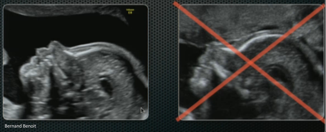
The successful 3D/4D ultrasound picture depends on the position of the fetus in the uterus and amount of amniotic fluid. It is not possible to get a 3D picture of the fetal face, if the fetal face is against the wall of the uterus or there is little placenta or amniotic fluid.
Monaco Professor Bernard Benoit from the Princess Grace Hospital made a summary of his work within 1 January 2007 – 20 October 2012, when his reception was visited by 8209 patients in different stages of pregnancy. During 1st trimester (within pregnancy week 11-14) the 3D picture of the fetal face succeeded in 86% of cases (2291 tests out of 2591), during 2nd trimester (within pregnancy week 14-28) in 92% of cases (2528 tests out of 2731) and during 3rd trimester (within pregnancy week 28-40) in 77% of cases (2240 tests out of 2887).
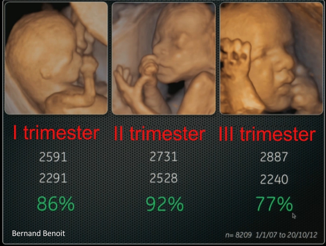
The highest probability to get a fetal face picture was within pregnancy weeks 14-28, when the fetal face appeared in 92% cases of ultrasound tests. Though, the fetal face was more true to nature after the 28th pregnancy week, but then it should be considered that every 4th visitor of the test will not see a fetal face during ultrasound.