Fetal ultrasound scan of the third trimester
During the third trimester of pregnancy the fetal ultrasound scan is performed with the help of the abdominal sensor within pregnancy weeks 34-36. At this stage of pregnancy the fetus has all vital organs fully developed and the period of fast growth and maturity continues. At this stage the fetus weighs 2400-2600 g and its length is 44-46 cm.
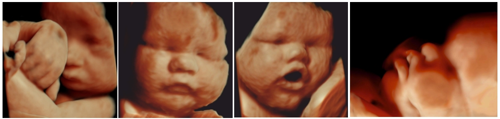
The fetus is already similar to a newborn infant as to the proportions of its body. It has bigger head compared to the body and thus most of them have already turned themselves upside down. It has developed mimic muscles of the face and it can smile. It opens its eyes. The connection has been created between the eye retina and brain and the child has a special glance in its eyes.
The pineal gland of the brain has started to produce a hormone called melatonin. Upon the impact of the hormone the fetus has developed its daily rhythm of life. Some fetuses are more active in the mornings, others in the evenings, but regardless of the latter the fetuses still sleep most of the time, i.e. 90-95% of the day.
The fetus has developed already all 5 senses at this stage of pregnancy
- It sees light gleaming through the mother’s abdominal covers
- It is able to hear the sounds coming outside the uterus, e.g. father’s voice
- It breaths and swallows amniotic fluid by feeling its smell and taste
- It has developed the skin tactile and pain sensitivity
Just as the newborn infant the fetus has already eyebrows and eyelashes in this stage of pregnancy. The toe and finger ends are covered with nails. Some fetuses have also hair. The heart beats 120-160 times a minute. This is twice as fast as mother’s, i.e. the total heartbeat of mother and father.
In the course of fetal ultrasound scan the position of the fetus is assessed whether the fetus is upside down or in breech position. If the fetus is in breech position, the external turn of the fetus into the position of head down can be offered to the woman in the hospital conditions which considerably increases the possibility for natural physiological labour and avoids the need of the Caesarean section.
In the course of ultrasound scan the size, maturity and position of placenta as to the cervix are assessed. It is essential to specify that the placenta and its blood vessels would not cover the entry of the cervix – this all for letting the child be born.
In case of previous Caesarean section the wholeness of the scar of the uterus is assessed and it would be observed whether the placenta covering the front wall of the uterus has not grown into the scar. This is essential information for the gynaecologist monitoring the pregnancy when preparing the birth management plan.
During ultrasound scan the amount of amniotic fluid is assessed, so that there would not be too little or too much of it. The amniotic fluid enables the fetus to move itself and as the fetus breaths and swallows amniotic fluid, the lungs and intestines are also developing.
By measuring the size of the skull, diameter of stomach and volume of thighbone, it is possible to assess the growth of the fetus and estimate the approximate birth weight of the child, knowing that the fetus gains 250-500 g with 2 weeks in this stage of pregnancy, i.e. maximum 1 kilogram within one month.
These mothers who have not gained enough weight at this stage of pregnancy and whose gain of stomach does not meet the size of pregnancy benefit from the assessment of fetal growth. The aim of the given ultrasound scan is to find on time these fetuses who have developed the intrauterine growth retention and whose growth dynamics and welfare condition need to be more often observed.
To assess the welfare condition of the fetus the ultrasound doppler scan is used in the course of which the bloodflow indices of fetal umbilical cord artery, brain arteries and intra-hepatic blood vessel ductus venosus is assessed. The mothers who do not clearly feel the movements of the fetus and who are worried about the welfare condition of the child also benefit from the fetal doppler scan.
Starting from the 36th pregnancy week it is possible to assess the probability of incurrence of late pre-eclampsia, by considering the values of blood pressure and placenta hormones of the mother with the bloodflow indicators of the uterine arteries received during ultrasound scan. This is very important information for the midwife monitoring the pregnancy for the due detection of pre-eclampsia before the aggravation of the child’s welfare condition for planning the induction of the labour.
If the premature induction of labour is planned (e.g. refractory diabetes during pregnancy, aggravating fetal growth retention, severe pre-eclampsia) the right timing of termination of pregnancy could benefit from the assessment of birth maturity of fetal lungs based on ultrasound scan.
In case of overcarriage starting from the pregnancy week 41 it is possible to estimate the start of spontaneous labour activities during the following week by measuring the length of the cervix. This is important information for the gynaecologist monitoring the pregnancy in assessing the need of induction of birth activities.
During the ultrasonic examination of foetal growth and wellbeing in the III. trimester, late preeclampsia can be assessed. This is very important as 75% of preeclampsia cases occur after the 37th week of pregnancy. This provides an opportunity to monitor women with a higher risk of preeclampsia more frequently and to detect the condition in a timely manner and prepare the child's lungs for the forthcoming birth.
At the end of the ultrasound scan the results of the ultrasound scan are explained to the family and, if needed, recommendations are given regarding the further monitoring of the fetus.
It is essential to know that most of the fetuses grow normally and are born healthy by creating a lot of joy for their parents in the future.