
Postimees published an opinion article by Dr Marek Šois entitled “Low Risk Is Not a Diagnosis”.
On 15 August 2026, Postimees' opinion portal published an article by Dr Marek Šois entitled “Low risk is not a diagnosis: pregnant women have the right to understand their options”.
The article focuses on the fundamental difference between prenatal screening and diagnostic testing. The OSCAR test and NIPT estimate the probability of certain chromosomal conditions, but they do not establish a diagnosis.
Amniocentesis, by contrast, allows analysis of fetal cells and provides a diagnostic result for the chromosomal or genetic variants covered by the selected laboratory test.[1,4,5]
The purpose of the article is not to favor one test over another or to recommend amniocentesis to every pregnant woman. It aims to help women understand what each test can answer, its capabilities and limitations, and what uncertainty remains even after a normal result.
Read the opinion article published in Postimees
Below, we publish an expanded version by the author that explains the differences between prenatal screening and diagnostic testing in greater detail.
Low risk is not a diagnosis: pregnant women have the right to understand their options
When a pregnant woman receives a low-risk screening result, she usually feels relieved. At the same time, questions may remain: Was the test accurate enough? Can the result be trusted? Does low risk mean that the baby does not have the condition being screened for?
These are important questions. Every pregnant woman deserves a clear explanation of what information a test actually provides and what cannot be concluded from its result.
What does “low risk” mean?
Prenatal screening does not establish a diagnosis; it estimates the probability that the condition being screened for is present.[4,5]
A low-risk result is good news, but it does not completely exclude the condition being screened for. Likewise, a high-risk result does not automatically mean that the fetus has a chromosomal or genetic condition.
All screening tests can produce both false-positive and false-negative results. A false-positive result indicates an increased risk even though the fetus does not have the variant in question. A false-negative result indicates low risk even though the variant is present.[4,5]
It is important to understand three principles:
-
the OSCAR test and NIPT estimate risk;
-
chorionic villus sampling and amniocentesis make diagnostic laboratory testing possible;
-
no normal test result can guarantee the birth of a completely healthy child.
What does the OSCAR test assess?
The OSCAR test combines a maternal blood test, an ultrasound examination and information about the mother's health. These are used primarily to estimate the fetal risk of Down syndrome, Edwards syndrome and Patau syndrome. The ultrasound examination also assesses early fetal development and anatomy, as well as possible signs of a chromosomal or genetic condition.
The OSCAR test is not merely a blood test or a measurement of nuchal translucency. A crucial part of it is a detailed ultrasound examination, which may identify developmental abnormalities even when the calculated risk of a chromosomal condition is low.
Nevertheless, the OSCAR test remains a screening test. A low-risk result does not mean that the fetal chromosomes have been examined directly.
Why is NIPT not a diagnostic test?
NIPT, or cell-free DNA analysis performed on a maternal blood sample, is the most accurate prenatal screening test for assessing the risk of the most common fetal chromosomal conditions, particularly Down syndrome, Edwards syndrome and Patau syndrome.[4]
People often mistakenly believe that NIPT analyses the baby's DNA directly. In reality, it analyses cell-free DNA fragments circulating in the mother's blood. The pregnancy-related fraction of these fragments originates predominantly from placental trophoblast cells.[4,10]
In most cases, the chromosomal make-up of the placenta and the fetus is the same. In rare cases, however, the placenta may contain a chromosomal variant that is not present in the fetus. The reverse is also possible: the variant may be present in the fetus but not reflected in the screening result as expected. This may be caused, for example, by confined placental mosaicism, a low proportion of pregnancy-related DNA, or other biological and technical factors.[4,10]
Therefore, even the most accurate and comprehensive NIPT remains a screening test. Increasing the number of conditions screened for does not make NIPT diagnostic.[4,6,7]
A high-risk NIPT result must be confirmed by diagnostic testing before making any irreversible decisions. An inconclusive NIPT result must not automatically be interpreted as low risk either. In such cases, the possible cause of the test failure must be assessed, and an individual plan for further investigation developed.[4,5]
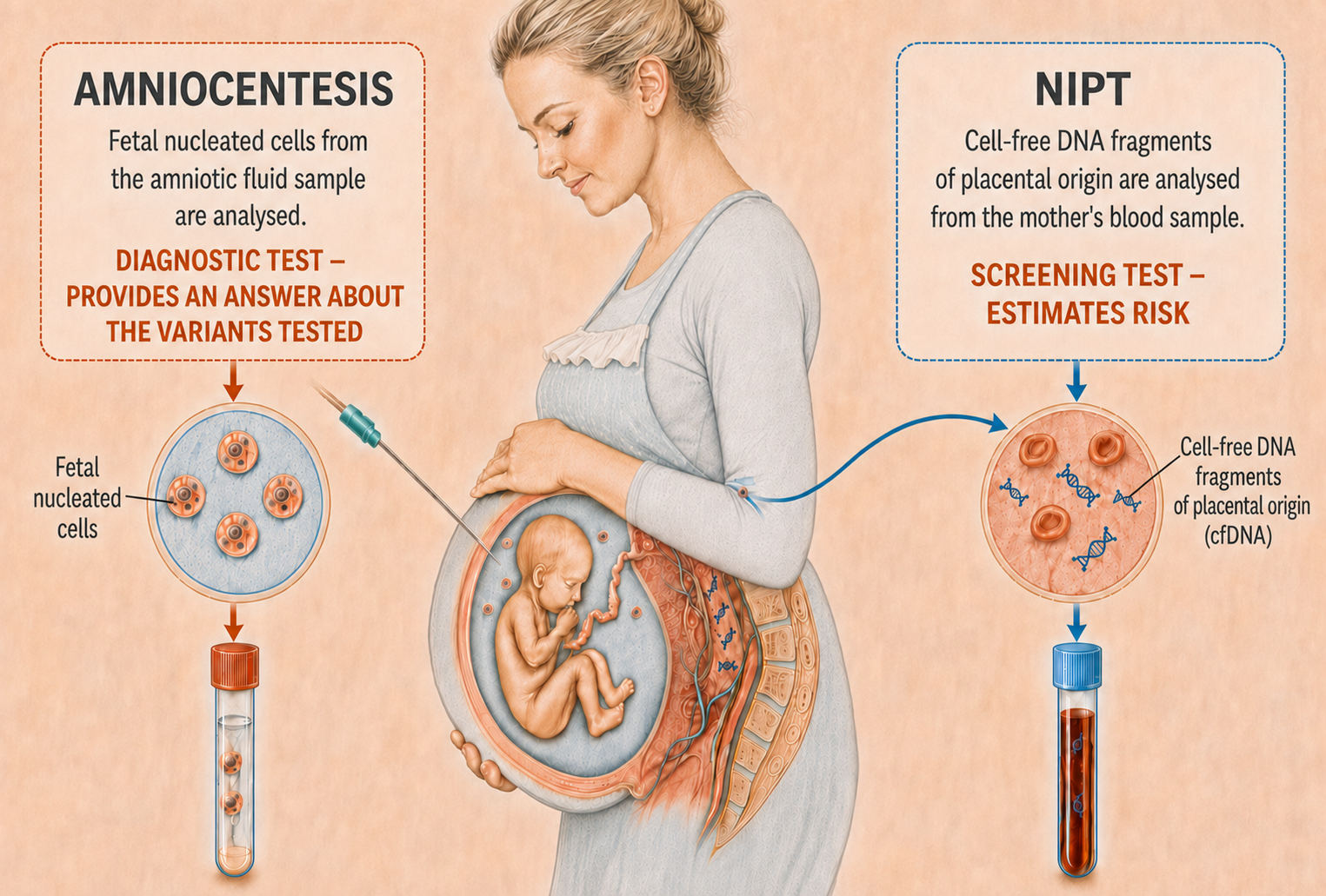
Illustration: NIPT mainly analyses cell-free DNA fragments of placental origin circulating in the mother's blood and estimates the risk of the variants being screened for. Diagnostic laboratory tests can be performed on fetal cells obtained through amniocentesis. Illustration: Marek Šois, created with the assistance of ChatGPT.
When a risk estimate is not enough
For many pregnant women and families, knowing that the risk of the chromosomal condition screened for is very low is sufficient. For some women, however, a diagnostic result for selected variants matters.
The question is not whether one choice is better than another. What matters is the kind of information a woman needs and how she views the different risks associated with testing.
In my view, the option of amniocentesis should not be discussed only when screening indicates a high risk. Following comprehensive counseling, a woman with a low-risk screening result also has the right to know that diagnostic testing is available.
International recommendations emphasize that, after appropriate counseling, patients have the right to accept or decline both prenatal screening and diagnostic testing.[4,5]
This does not mean that every pregnant woman should undergo amniocentesis. It means women must be given enough information to decide whether a risk estimate is sufficient or whether they want a diagnostic result for selected chromosomal or genetic variants.
Studies from Israel have shown that clinically significant chromosomal variants may be present even when screening indicates low risk and ultrasound findings are normal, and that these variants may not be detected by NIPT.[6–8] However, these findings cannot be applied directly to the entire pregnant population because the studies involved women who had already chosen invasive testing.
A 2025 Dutch study of 46,007 pregnancies with normal cfDNA screening results estimated the residual risk of a pathogenic chromosomal aberration at approximately 0.14–0.15%. When an ultrasound abnormality was identified in the fetus, the residual risk increased substantially.[9]
Numerical results vary between studies because study populations, NIPT methods, and follow-up duration differ. The shared conclusion is nevertheless clear: a low-risk NIPT result reduces the probability of the variants screened for, but it does not reduce the risk of every possible chromosomal abnormality to zero.[6–9]
Why does amniocentesis begin with an ultrasound examination?
When people think of amniocentesis, they often think first of the needle. For a fetal medicine specialist, however, the examination begins with a detailed fetal ultrasound.
At approximately 16 weeks of pregnancy, many fetal anatomical structures can already be assessed and signs that may indicate a chromosomal or genetic condition can be sought. This examination does not replace the later fetal anatomy scan, which is usually performed at 20–21 weeks of pregnancy.
If the ultrasound reveals a developmental abnormality or a sign suggestive of a genetic condition, this may influence the choice of laboratory test. In such cases, it may be necessary to involve a clinical geneticist and, in addition to chromosome analysis, to examine a specific gene or group of genes.
Even when the ultrasound findings are normal, a woman may still choose diagnostic testing after counseling if obtaining the most definitive possible answer about selected variants is important to her.[4,6–9]
What is analyzed from the amniotic fluid?
During amniocentesis, a small amount of fluid is taken from the amniotic sac surrounding the fetus under continuous ultrasound guidance. The amniotic fluid contains fetal cells that can be used for different laboratory analyses.[1,3]
The choice of test depends on the clinical circumstances and the patient's information needs. Possible analyses include, for example:
-
rapid testing for the most common changes in chromosome number;
-
chromosomal microarray analysis, also known as submicroscopic chromosome analysis;
-
targeted testing for a specific chromosomal or genetic variant;
-
broader genetic testing when medically indicated.
Chromosomal microarray analysis can detect small missing or additional DNA segments that conventional chromosome analysis or NIPT targeting the most common trisomies may not identify.[6–9]
In rare cases, however, the analysis may identify a variant whose implications for the child's future health are not clear. For some findings, it is not possible to predict precisely whether the condition will manifest, when it will, or how severely it will affect the child. Therefore, discuss the analysis's capabilities and limitations before testing.[11,12]
What risks are associated with amniocentesis?
Amniocentesis is an invasive procedure. According to current evidence, the additional risk of miscarriage associated with a procedure performed by an experienced specialist is approximately 0.1%, or around 1 in 1,000.[2]
In a 2019 systematic review and meta-analysis, the additional procedure-related risk of miscarriage was 0.12% when groups with comparable baseline risks were analyzed. The risk is not zero, and an exact individual risk cannot be guaranteed in advance.[2]
This risk must neither be minimized nor concealed. At the same time, women must be able to weigh it against the information that diagnostic testing may provide.
Amniocentesis should not be performed before 15 weeks of pregnancy. In practice, 16–18 weeks is often a suitable time for the procedure. A suitably trained specialist must perform it under continuous ultrasound guidance.[1,3]
Diagnostic testing cannot answer every question either
Amniocentesis is not a single laboratory test with a fixed scope. The content of the diagnostic result depends on which analysis is ordered on the amniotic fluid sample.
A normal chromosomal microarray result does not exclude most conditions caused by single-gene variants. Even broader genetic testing cannot detect every disease-causing variant or predict the child's entire future health and development.
Likewise, a normal ultrasound examination does not exclude every developmental abnormality. Some conditions may become apparent only later in pregnancy, after birth, or as the child grows.
Therefore, no doctor can honestly promise that a normal test result guarantees a healthy birth. The doctor's task is to explain:
-
what the selected test can detect;
-
what the test cannot detect;
-
how reliable the result is;
-
what uncertainty remains;
-
what will be done if an abnormality or a variant of uncertain significance is identified.[4,11,12]
A diagnostic service must cover the entire care pathway
Amniocentesis should not be limited to performing the procedure and sending a sample to the laboratory.
Comprehensive care includes:
-
a detailed ultrasound examination;
-
assessment of the patient's previous test results;
-
pre-test counseling;
-
selection of the appropriate laboratory analysis;
-
amniocentesis under continuous ultrasound guidance;
-
a clear explanation of the laboratory result;
-
involvement of a clinical geneticist and other specialists when necessary;
-
development of a plan for further monitoring, delivery or treatment.[1,3–5]
A laboratory report alone is not enough for the patient. She must understand what an identified or absent variant means for her child and what the next steps are.
A diagnosis does not decide for a woman
The purpose of prenatal diagnosis is not to decide which child deserves to be born. A diagnosis does not oblige a woman to terminate a pregnancy, and a low-risk screening result does not oblige her to accept a level of uncertainty that she finds unacceptable.
A woman has the right to hope for the birth of a healthy child and to use the testing options available before birth. Equally, she has the right to continue the pregnancy and give birth even when the fetus has been diagnosed with a condition or developmental difference. But such a decision must be informed.
If a fetal condition is identified, the family should receive the clearest possible answers to the following questions:
-
what the diagnosis means for the child;
-
how certain the diagnosis is;
-
what the severity and prognosis may be;
-
whether the condition can be treated during pregnancy or after birth;
-
what treatment, care or additional support the child may need;
-
whether delivery should be planned at a hospital capable of providing specialist care;
-
how the child's condition may affect the future life of the rest of the family;
-
what options the family has if they wish to continue the pregnancy;
-
what options are permitted by law if the woman does not wish to continue the pregnancy after receiving a serious diagnosis.
Knowing the diagnosis is not only about choosing whether to continue or terminate the pregnancy. It can give the family time to understand the situation, prepare emotionally and practically, find the specialists they need, and organize treatment and support for the child after birth. For some conditions, a prenatal diagnosis may also directly influence the timing, mode and place of delivery.
Continuing a pregnancy after a fetal condition has been diagnosed is an informed decision worthy of respect. Equally, no one should be compelled to continue a pregnancy in a situation where testing for a serious condition was available, but the woman was not told about it.
A doctor must neither direct a woman towards terminating a pregnancy nor urge her to continue it at any cost. But a doctor must not deprive her of the opportunity to know that diagnostic testing is available.
The doctor's most important responsibility
My role is not to decide which child a family is able or willing to live with. My role is to help a woman understand what can be known before birth, what cannot be known, and what choices and possible consequences follow from the result.
After counseling, one woman may decide that screening is sufficient for her. Another may want diagnostic testing. Both decisions can be reasonable when made after honest, understandable counseling and reflect the values and information needs of the woman or family.[5,11]
The most unjust situation arises when a woman learns only after her child is born that the condition could have been investigated during pregnancy, but she was never told that this option existed.
Lack of knowledge is not an informed choice when a woman has not been offered the relevant information or the option of diagnostic testing.
A doctor should therefore not decide on a woman's behalf that a low-risk screening result must be sufficient for her. She must be given clear information and the right to decide how much certainty she needs and what degree of uncertainty she is prepared to live with.[4,5,11,12]
Screening provides a probability. Diagnostic testing may provide an answer. The decision belongs to the woman.
Read the opinion article published in Postimees
Author: Dr Marek Šois
Obstetrician-gynecologist specializing in fetal medicine
Cover image caption: Dr Marek Šois explaining the possibilities and limitations of prenatal testing to an expectant family. Photograph: Kristiina Männik.
References
-
Ghi T, Sotiriadis A, Calda P, Da Silva Costa F, Raine-Fenning N, Alfirevic Z, et al.; International Society of Ultrasound in Obstetrics and Gynecology. ISUOG Practice Guidelines: invasive procedures for prenatal diagnosis. Ultrasound Obstet Gynecol. 2016;48(2):256–268. doi:10.1002/uog.15945
-
Salomon LJ, Sotiriadis A, Wulff CB, Odibo A, Akolekar R. Risk of miscarriage following amniocentesis or chorionic villus sampling: systematic review of literature and updated meta-analysis. Ultrasound Obstet Gynecol. 2019;54(4):442–451. doi:10.1002/uog.20353
-
Royal College of Obstetricians and Gynaecologists. Amniocentesis and Chorionic Villus Sampling. Green-top Guideline No. 8. London: RCOG; 2021. Reviewed December 2024. RCOG guideline
-
Society for Maternal-Fetal Medicine; Rink BD, Dugoff L, Kuller JA; SMFM Publications Committee. Society for Maternal-Fetal Medicine Consult Series #74: Cell-free DNA screening for aneuploidies: updated guidance. Pregnancy. 2025;1(6). doi:10.1002/pmf2.70139
-
Royal College of Obstetricians and Gynaecologists. Supporting women and their partners through prenatal screening for Down’s syndrome, Edwards’ syndrome and Patau’s syndrome. London: RCOG; 2020. RCOG guidance
-
Maya I, Salzer Sheelo L, Brabbing-Goldstein D, Matar R, Kahana S, Agmon-Fishman I, et al. Residual risk for clinically significant copy number variants in low-risk pregnancies following exclusion of noninvasive prenatal screening-detectable findings. Am J Obstet Gynecol. 2022;226(4):562.e1–562.e8. doi:10.1016/j.ajog.2021.11.016
-
Maya I, Salzer Sheelo L, Brabbing-Goldstein D, Matar R, Kahana S, Agmon-Fishman I, et al. Clinical utility of expanded non-invasive prenatal screening compared with chromosomal microarray analysis in over 8000 pregnancies without major structural anomaly. Ultrasound Obstet Gynecol. 2023;61(6):698–704. doi:10.1002/uog.26177
-
Sagi-Dain L, Salzer Sheelo L, Brabbing-Goldstein D, Matar R, Kahana S, Agmon-Fishman I, et al. Prevalence of high-penetrant copy number variants in 7734 low-risk pregnancies. Am J Obstet Gynecol MFM. 2023;5(12):101201. doi:10.1016/j.ajogmf.2023.101201
-
Iglesias AI, Van Opstal D, Thurik FF, Drost M, Weerts MJA, Joosten M, et al. Residual risks of fetal chromosome aberrations when cell-free DNA prenatal screening is normal: a retrospective study. Prenat Diagn. 2025;45(12):1559–1571. doi:10.1002/pd.6888
-
Van Opstal D, van Veen S, Joosten M, Diderich KEM, Govaerts LCP, Polak J, et al. Placental studies elucidate discrepancies between NIPT showing a structural chromosome aberration and a differently abnormal fetal karyotype. Prenat Diagn. 2019;39(11):1016–1025. doi:10.1002/pd.5531
-
Hochner H, Daum H, Douiev L, Zvi N, Frumkin A, Macarov M, et al. Information women choose to receive about prenatal chromosomal microarray analysis. Obstet Gynecol. 2020;135(1):149–157. doi:10.1097/AOG.0000000000003610
-
Brabbing-Goldstein D, Reches A, Svirsky R, Bar-Shira A, Yaron Y. Dilemmas in genetic counseling for low-penetrance neuro-susceptibility loci detected on prenatal chromosomal microarray analysis. Am J Obstet Gynecol. 2018;218(2):247.e1–247.e12. doi:10.1016/j.ajog.2017.11.559