Loote Ultrahelikeskus offers early fetal anatomy scan of I trimester within OSCAR test
Dr. Marek Šois passed this year continuing training programme carried out by the London School of Ultrasound in Harpenden, England. This programme was focused on diagnosing pathologies and syndromes to be detected within 12-13 weeks of pregnancy at the fetal early ultrasound scan and improvement of technical skills of the latter.
The improvements were carried out by Dr. Fred Usakov, internationally recognized specialist of fetal medicine and echocardiography, who is also an expert consultant of University College London Hospital and a member of the committee of continuing training of International Society of Ultrasound in Obstetrics and Gynecology (ISUOG).
The continuing training programme focused on the early ultrasound scan during 11-13 weeks of pregnancy within which approximately 100 fetal anomalies and syndromes were examined.
London School of Ultrasound
- 02.02.-04.02.2018
“Fetal heart: introduction and essential echocardiography.“ - 09.03.-11.03.2018
“Early neurosonography: brain, spine and face.“ - 11.05.-13.05.2018
“Fetal echo: state of the art.“ - 15.06.-17.06.2018
“Important fetal anomalies.“ - 03.08.-06.08.2018
“Fetal cardiology for experts.“ - 13.08.-16.08.2018
“Early fetal scan conference 2018.“
The international study conducted during 1991-2014 found that it is possible to detect approximately half of the congenital fetal abnormalities at the ultrasound examination of the first trimester (1).
It is widely incorrectly understood that the prenatal scan of Down syndrome offered as the national programme is equal to the ultrasound scan of I trimester provided in Loote Ultrahelikeskus.
- The percentage of the fetuses with Down syndrome of the congenital development abnormalities is only 10%. (2)
- The rate of children with Down syndrome among the children with mental disabilities is only 10%. (3)
- The best test for detection of fetuses with Down syndrome is NIPT test (Panorama test), not the ultrasound scan (OSCAR test). (4)
- More than 90% of the congenital structural abnormalities and about 90% of the children with mental disabilities remain undetected in performing NIPT test (Panorama test).
Loote Ultrahelikeskus wishes to achieve new objectives at the ultrasound scan of 12-13 weeks of pregnancy – to focus on the early fetal anatomy and detection of fetuses with congenital abnormalities, not to seek only for ultrasound markers characteristic of chromosomal diseases and by dividing the fetuses based on the latter either to the low or high risk group of chromosomal disorders.
The samples on the severe abnormalities to be potentially detected during the early fetal ultrasound scan:
- Congenital heart defects
- Spina bifida
- Congenital umbilical cord hernia
- Lethal skeletal dysplasias
- Limb defects
- Gastroschisis
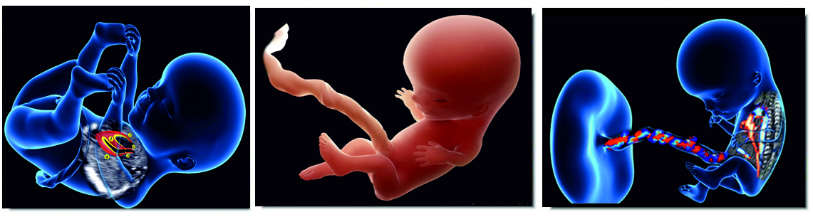
The aim of the ultrasound scan of the first trimester is the screening of congenital abnormalities within which the fetal body systems are systematically examined from toe to head by paying greater attention to the assessment of fetal heart and brain structures. The best time for the screening of the first trimester is 12-13 weeks of pregnancy (80% of OSCAR tests are conducted in this period in Loote Ultrahelikeskus). Before the 12th week of pregnancy, the fetal body structures are complicated to assess due to fetal immaturity (foremost fetal brain) and insufficient resolution of an ultrasound scan.
Why does Loote Ultrahelikeskus recommend the assessment of early fetal body structures during 11-13 weeks of pregnancy?
- This period enables to detect half of the abnormalities potentially.
- It is possible to detect most of the severe abnormalities with the worst prognosis.
- Combining ultrasound scan with Panorama test is possible.
- It is possible to carry out early diagnostic test – chorionic biopsy, rather than waiting up to 15-16 weeks of pregnancy for the conduct of the amniotic fluid test.
- As the fetus is close to the cervix, it is possible to use a vaginal ultrasound scan to specify the diagnosis.
- Some congenital abnormalities are more detected and thus easier to be found at the early ultrasound scan.
- Under the ultrasound scan, there is still time for more precise diagnosis (karyotype, submicroscopic analysis, the study of the whole genome, pre-natal infection analyses) and consultation with colleagues (second opinion from the colleagues, geneticist, pediatric surgeon, pediatric cardiologist).
- If required by the family, it is possible to terminate the pregnancy in case of severe fetal abnormalities in a more safe way.
- In case of some abnormalities, it is possible to monitor the development of fetal malformations in time during 16-20 pregnancy weeks to assess the progress of the disease and give a more specific prognosis on the child’s life and health based on the latter.
The screening of congenital heart failures should be started already during the first trimester of pregnancy, as:
- The congenital heart defects are more frequent congenital abnormalities (about 1: 100 of the newborn children have a congenital heart defect with different degree of severity).
- These are mostly severe abnormalities (most of the congenital heart failures require operational treatment).
- There are numerous forms of congenital heart failures (approximately 200 different heart failures).
- The congenital heart defects of the newborn cause a devastating impact on the whole family.
- The congenital heart defects are often combined with other fetal abnormalities (in about 40% of cases).
- The congenital heart defects are complicated to detect at the general scan (only 50% of the congenital heart defects are pre-natally identified each year in England).
- Most of the congenital heart defects can be diagnosed already within 12-13 weeks of pregnancy (85% of the severe heart failures can be diagnosed in the course of early ultrasound scan).
- The results of a pre-natal scan of congenital heart defects are considerably better when the combined pre-natal scan of heart failures is performed (during 12th and 20th weeks of pregnancy).
- The prenatal detection of congenital heart defects improves the survival rate and life quality of children.
Used literature:
- Karim JN, Roberts NW, Salomon LJ, Papageorghiou AT. A systematic review of first-trimester ultrasound screening for detection of fetal structural anomalies and factors that affect screening performance. Ultrasound Obstet Gynecol. 2017 Oct;50(4):429-441
- http://www.eurocat-network.eu/accessprevalencedata/prevalencetables
- Acharya K. Prenatal testing for intellectual disability: misperceptions and reality with lessons from Down syndrome. Dev Disabil Res Rev. 2011;17(1):27-31
- Gil MM, Accurti V, Santacruz B, Plana MN, Nicolaides KH. Analysis of cell-free DNA in maternal blood in screening for aneuploidies: an updated meta-analysis. Ultrasound Obstet Gynecol. 2017 Sep;50(3):302-314