Loote Ultrahelikeskus offers fetal neurosonography for detection of disorders of central nervous system and specification of diagnosis
Certificate of attendence Fetal Neurosonography Mastercalss
Dr. Marek Šois passed theoretical and practical training for fetal neurosonography in Sourasky Medical Center in Tel Aviv, Israel, by professor Gustavo Malinger, one of the world’s most recognized fetal neurosonographer.
The aim of fetal neurosonography is to assess the structures of fetal nervous systems. The ultrasound examination includes the assessment of the fetal brain as well as spine. The given ultrasound examination can be performed also at the existence of the indications at the end of the first trimester and at the beginning of the second trimester during 13-16 weeks of pregnancy, but the best time for the latter is still in the second trimester after the 20th pregnancy week. As the standard finding in the anatomical screening does not exclude the incurrence of brain damage in the third trimester the test should be repeated at certain indications during pregnancy weeks 28-32.
The fetal neurosonography is carried out, if required, as combined with abdominal ultrasound probe as well as with vaginal ultrasound probe depending on the position of the fetus and peculiarity of the pathology under examination.
Indications for neurosonography
- a close relative having a child with psychomotor retardation or mental disability
- suspicion of pathology of fetal brain or spine incurred in the fetal anatomic screening test
- expansion of fetal brain ventricles
- cyst or its suspicion in the fetal brain
- non-conformity of fetal cranium with the size of pregnancy (too small or big cranium)
- existence of uterine infections (cytoplasmosis, toxoplasmosis)
- severe fetal growth retention
- monochorionic twin pregnancy
- inborn development disorder in other organic systems (developmental abnormalities of heart, clubfoot, deformations of limbs, developmental abnormalities of kidneys, cleft upper lip, hernia of umbilical cord etc).
At the detection of pathology of fetal central nervous system the family is referred to the consultation of geneticist and children’s neurologist, if necessary. In certain cases the patient is referred to MRT (magnetic resonance tomography) test for specifying the diagnosis and implementation of pregnancy management plan.
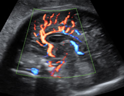
Pericallosal artery