Gynaecological 3D ultrasound scan
3D ultrasound is often more detailed than a standard 2D examination and allows the physician to evaluate the uterus from multiple sections and perspectives simultaneously. A 3D ultrasound examination of the uterus is primarily performed to obtain a more precise spatial (three-dimensional) view of its shape and structure. It is used for both diagnostic purposes and to уточнить the treatment plan.
Main indications and purposes:
-
More accurate detection of congenital uterine anomalies (septate uterus, bicornuate uterus, arcuate uterus);
-
Evaluation of the causes of infertility and recurrent miscarriages;
-
Assessment of uterine cavity deformities (polyps, fibroids, adhesions);
-
Improved visualisation of endometriosis and adenomyosis;
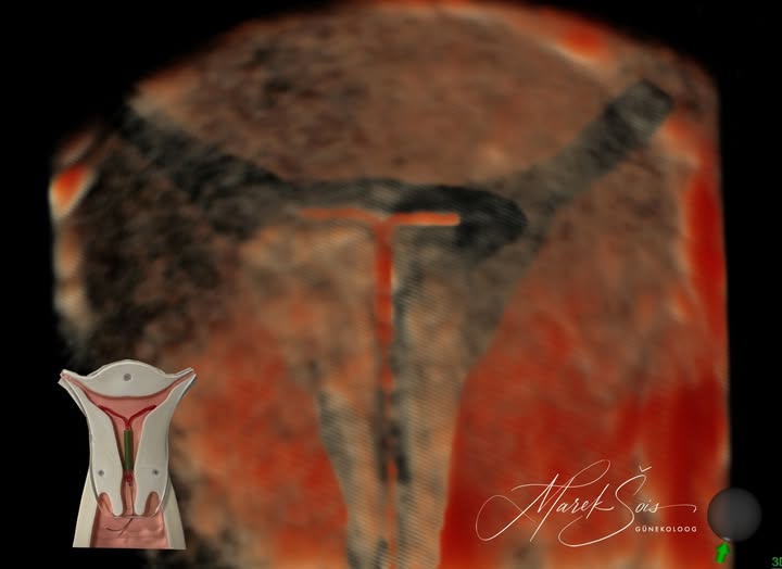
-
Checking the position of an intrauterine device (IUD);
-
Planning surgical procedures or fertility treatment and evaluating outcomes.