The Fetal Ultrasound Center received a long-awaited chorionic villus sampling simulator
Chorionic villus sampling is an invasive prenatal diagnostic test in which tissue material is taken from the developing placenta (chorion) under ultrasound control. The aim of the procedure is to detect possible chromosomal and genetic diseases in the fetus before birth.
Dr. Marek Šois. Chorionic villus sampling for a placenta located on the back wall of the uterus
This is an invasive study, in which there is a 1:500 chance of miscarriage per procedure. The greater the experience of the physician performing the chorionic villus sampling, the lower the risk to the life and health of the fetus.
It is not always possible to perform a chorionic villus sampling for technical reasons. In such cases, after 4 weeks of gestation, the woman is offered an amniotic fluid test at the 16th week of her pregnancy, in which material is taken from the amniotic fluid surrounding the baby - instead of the placenta - for chromosomal and genetic testing. The most common reason why the physician fails to perform a chorionic biopsy is a uterus located on the posterior wall of the placenta.
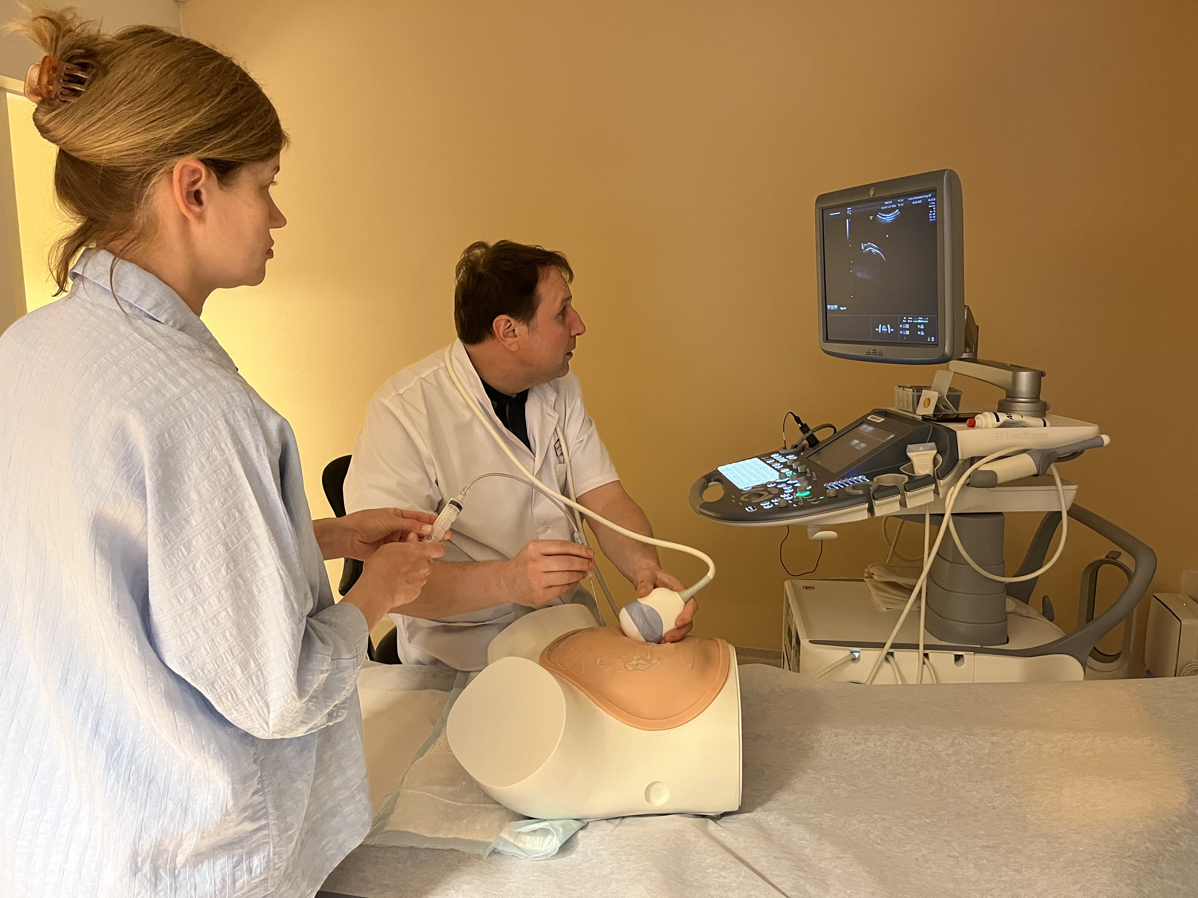
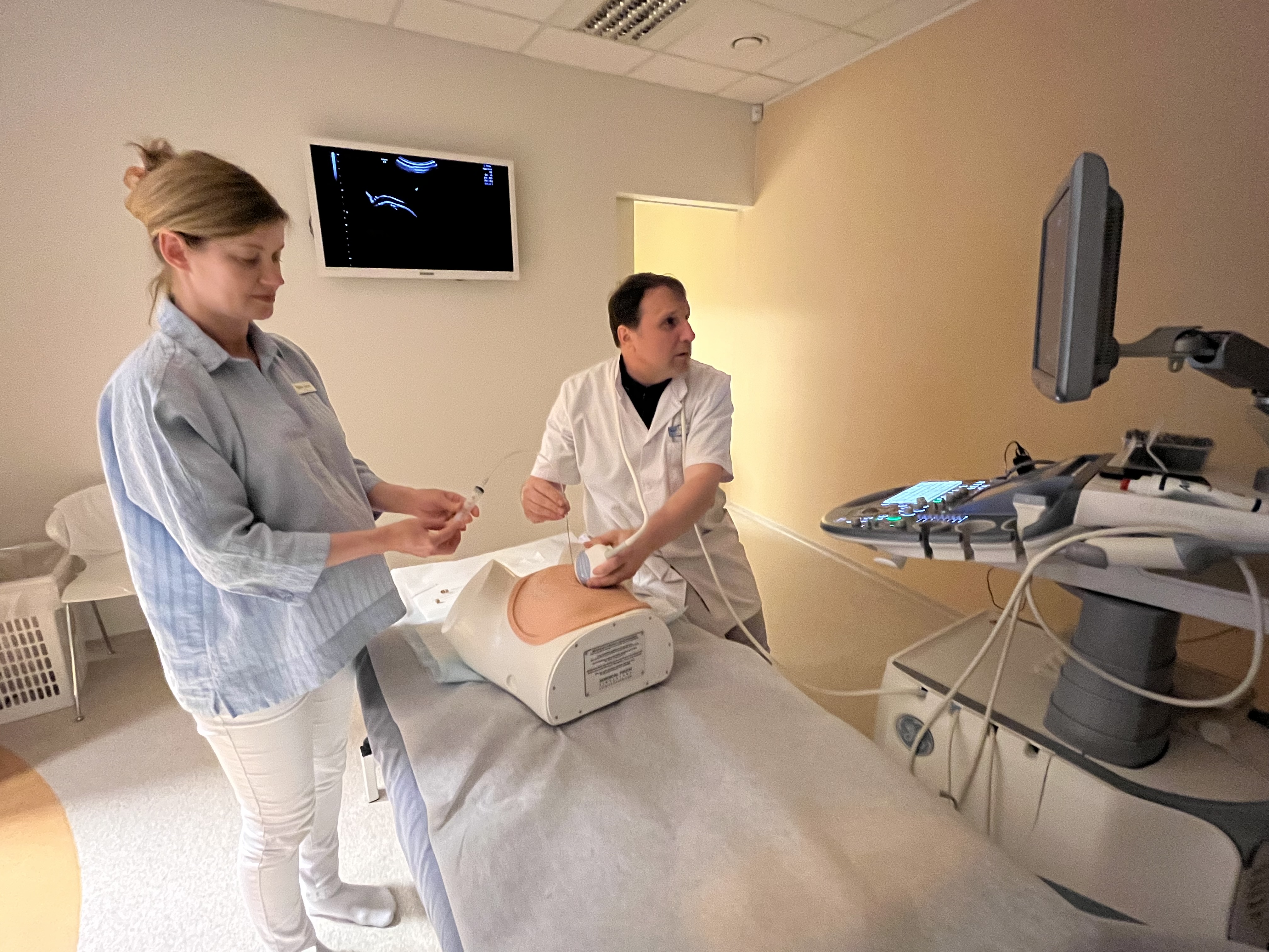
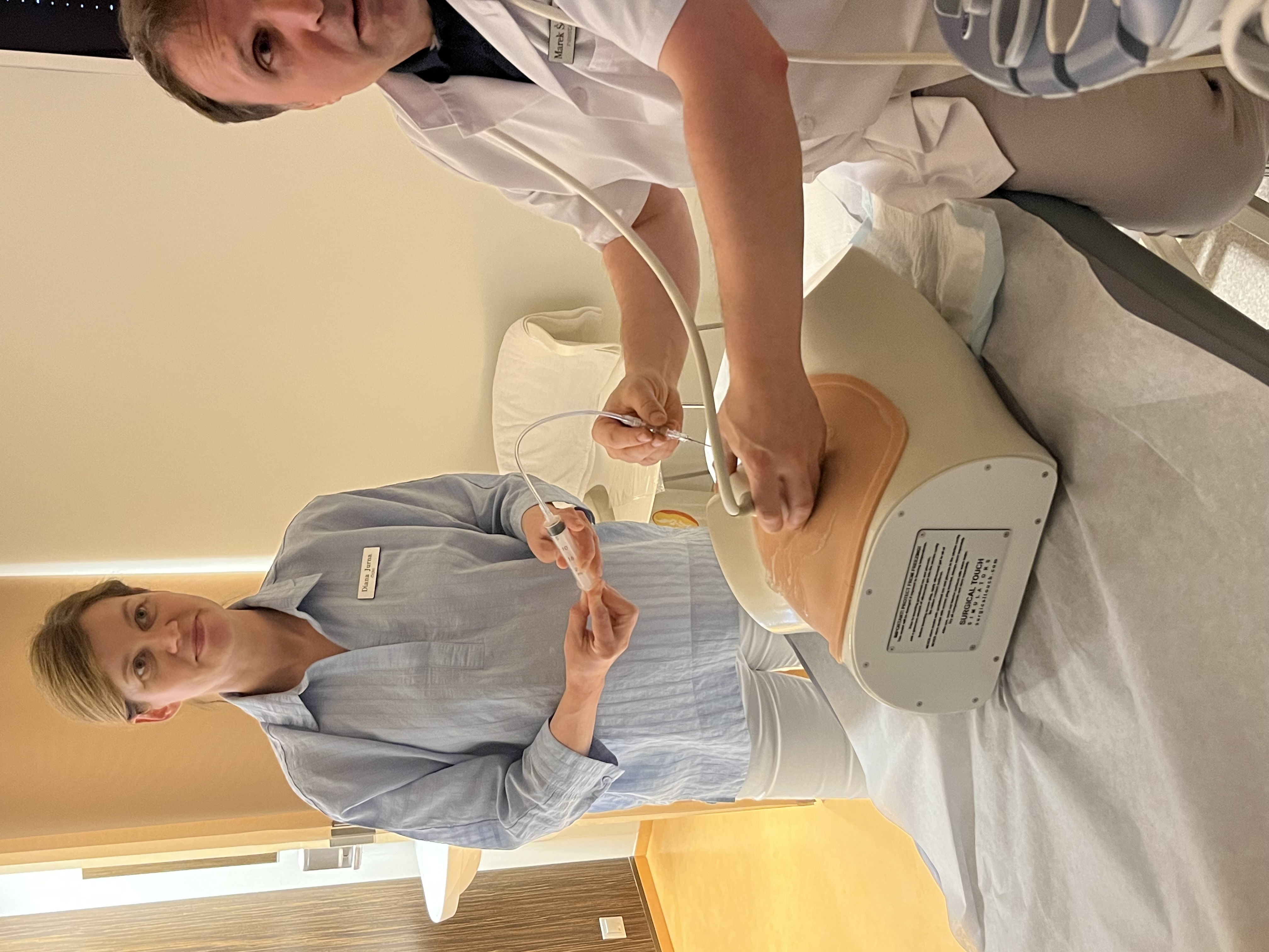
Francis LeBouthillier of Surgical Touch in Canada handcrafted a special simulator for Dr. Marek Šois in 7 months, to provide a tool for learning how to perform a chorionic villus sampling.
The chorionic villus sampling simulator consists of an imitation of a woman's lower body with a uterus of 13 gestation weeks in size, in which a latex reservoir filled with an amniotic fluid can be placed, along with an artificial placenta. The simulator is covered with a multilayer silicone coating imitating the skin, which mimics the various anatomical layers of the female abdomen. The artificial uterus is located in a small pelvis in a gel medium, which can be studied with an ultrasonic transducer through the simulator's abdominal covers. The aim of the simulator is to learn how to accurately target placental tissue by puncturing the abdomen with a biopsy needle in different placental positions without damaging the amniotic membrane.
After learning for several weeks on the simulator, Dr. Marek Šois successfully performed a chorionic villus sampling using the technique of Professor Tak Yeung Leung from Hong Kong on a woman whose uterus was located on the posterior wall of the placenta.
The OSCAR test conducted at the Fetal Ultrasound Center revealed that the 69 mm (13 weeks) long fetus had a very large nuchal translucency (NT was 6.77 mm). In addition to congenital heart defects, several serious chromosomal diseases or genetic defects may be the cause of an increased occipital fold. A detailed early ultrasound of the fetal heart showed that the heart was healthy, and the rapid response to the chorionic villus sampling, which came the next day, was also fine. The geneticists also performed a final chromosomal analysis of the placental material, and after two weeks it was confirmed that the fetus does not have any of the common chromosomal or genetic diseases. To the woman's great relief, she did not have to wait another 4 weeks for an amniocentesis, but instead received a quick final response thanks to the success of the chorionic biopsy.
Dr. Marek Šois is extremely pleased with the opportunity to practice on this unique chorionic villus sampling simulator – it gives him a greater confidence that he can help his patients by performing a chorionic villus sampling, even if performing it may be technically difficult.
 |
 |
 |
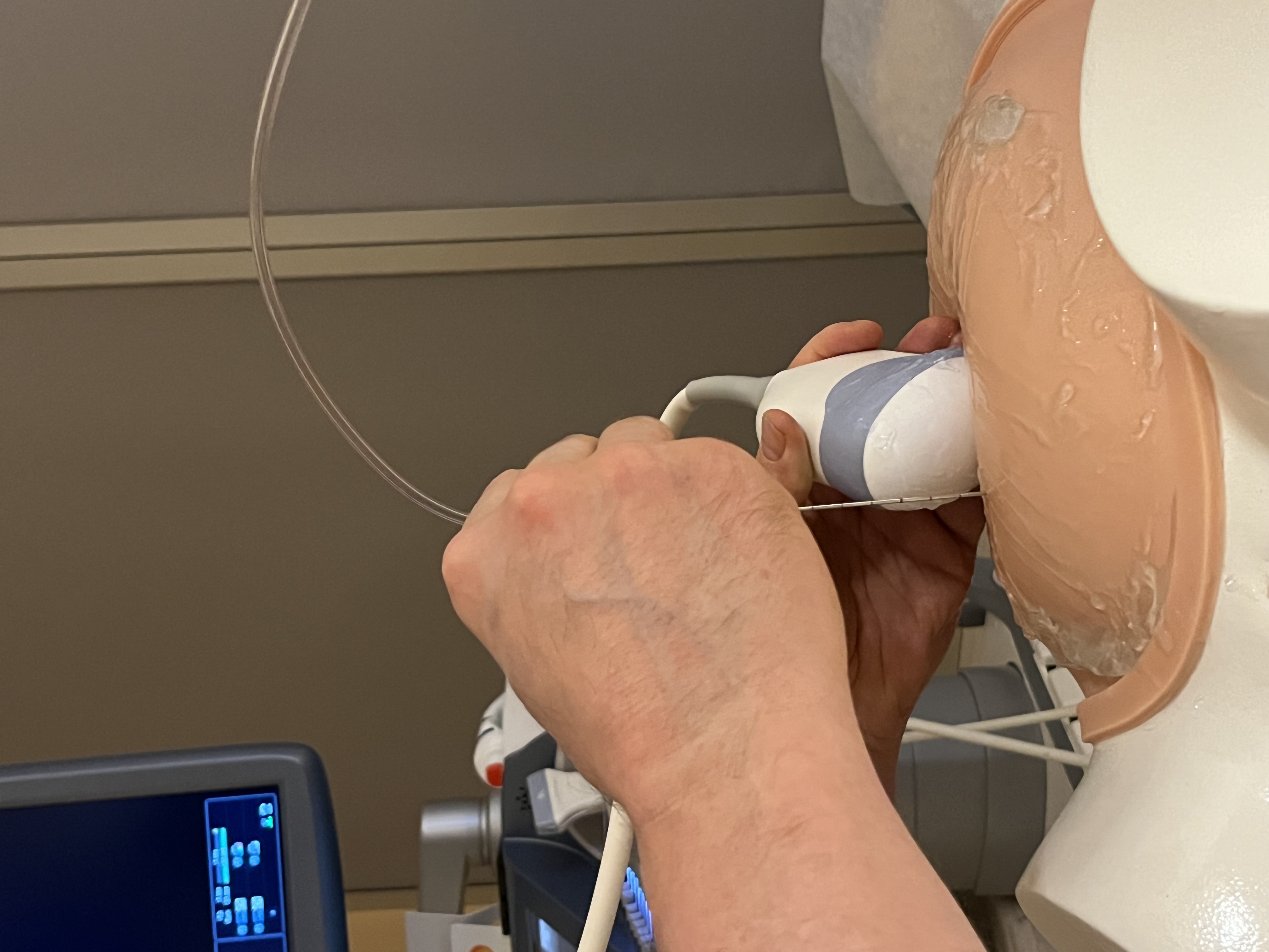 |
Training on a chorionic villus sampling simulator